15 Mar The Role Of Physical Therapy In Anal Fissure Rehabilitation The Role Of Physical Therapy In Anal Fissure Rehabilitation Read More
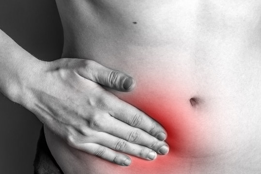
11 Mar Understanding The Appendix: What You Need To Know Understanding The Appendix: What You Need To Know Read More
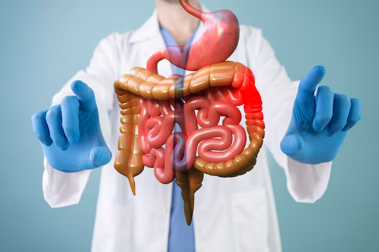
08 Mar Emerging Technologies And Innovations In Hernia Treatment Emerging Technologies And Innovations In Hernia Treatment Read More
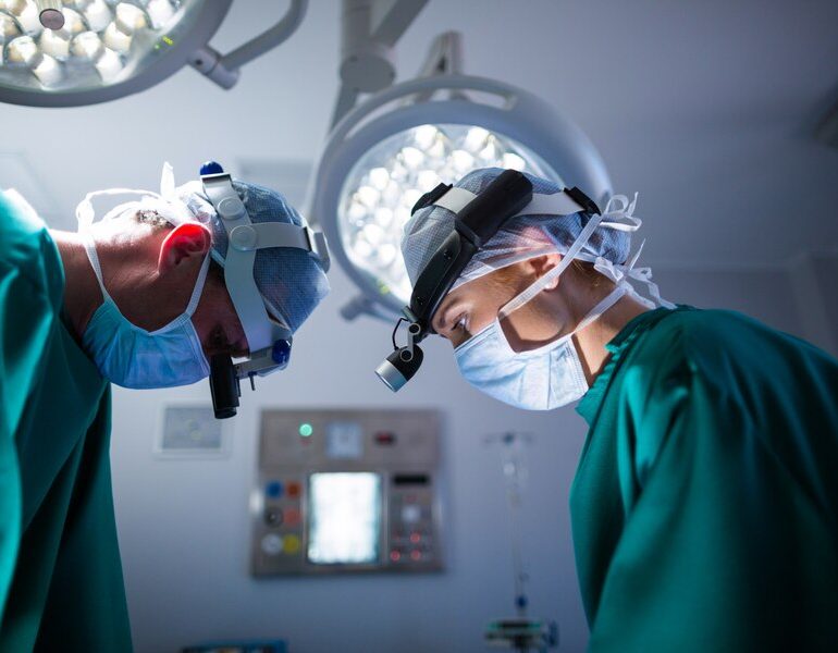
05 Mar Exploring The Latest Techniques In Laparoscopic Surgery Exploring The Latest Techniques In Laparoscopic Surgery Read More
29 Feb Circumcision And HIV Prevention: Examining The Latest Research Findings Circumcision And HIV Prevention: Examining The Latest Research Findings Read More
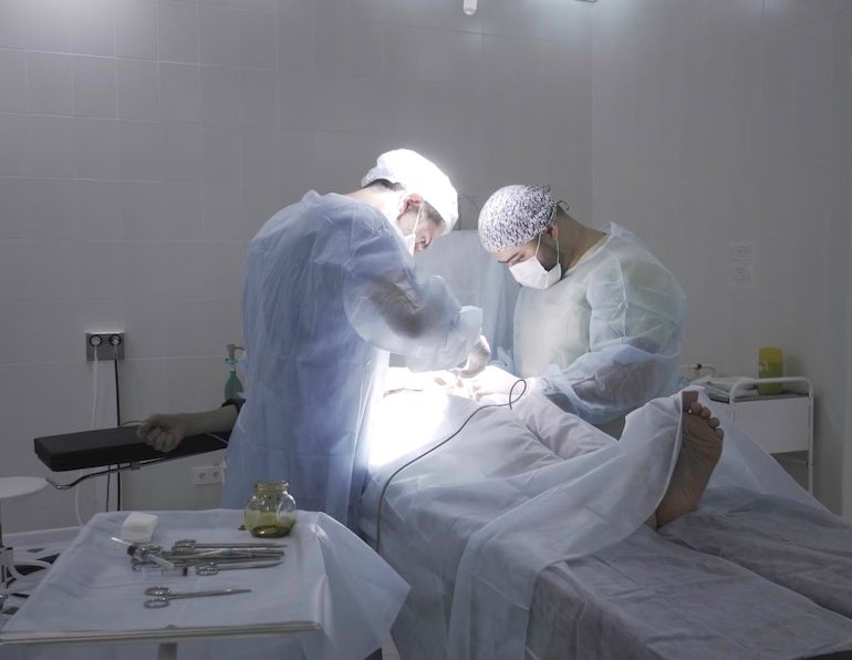
27 Feb Postoperative Care And Recovery After Gastrointestinal Surgery Postoperative Care And Recovery After Gastrointestinal Surgery Read More
26 Feb Innovative Approaches And Emerging Therapies In Pilonidal Sinus Management Innovative Approaches And Emerging Therapies In Pilonidal Sinus Management Read More
23 Feb Healing The Cracks: Innovative Approaches To Anal Fissure Treatment Healing The Cracks: Innovative Approaches To Anal Fissure Treatment Read More
22 Feb Complications Of Untreated Piles: What You Need To Know Complications Of Untreated Piles: What You Need To Know Read More